flowchart TD
  A[Nose] -- smell --> B["Cribriform plate"]
  B --> C["Olfactory Sulci"]
  C -- Skips Thalamus --> D["Olfactory Tract"]
  D --> E[Olfactory Bulb]

  E --> F[Insula gustation but i wrote that this was wrong]
  E --> G[Piriform Cortex]
  E --> H[Amygdala]
  E --> I[Entorhinal Cortex]

  F --> J{Gustation}
  G --> K{Olfaction processing}
  H --> L{Emotion of Fear}
  I --> M{Formation of Memory}
CN I: Olfactory nerve
- Type: Sensory
- Function: Sense of smell
- Dysfunction: Anosmia (loss of smell)
Pathway:
First order neurons:
- Smell is detected by specialized chemoreceptors on bipolar primary sensory neurons found in the olfactory neuroepithelium1
- Olfactory nerve (made up of axons combined with other receptor cell axons)1
- Pass through Cribriform plate of Ethmoid bone1
- Terminate on the olfactory bulb (main relay station of olfactory pathway)1
Second order neurons:
- Olfactory tract
- Split: Medial & Lateral striae
- Medial Stria: Projects to subcallosal gyrus
- Lateral Stria: continues on to Parahippocampal gyrus
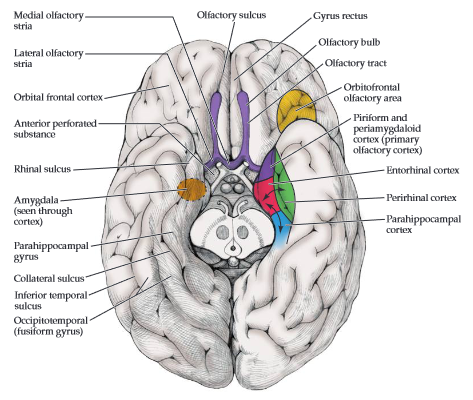
Relevant Structures
The olfactory nerve does not have peripheral ganglia
Olfactory cortex: no single structure - Piriform cortex: - Located: Below lateral olfactory stria - Function: Olfactory processing - Amygdala: - Located: Anterior to Temporal/Inferior horn of Lateral Ventricle - Function: Emotion & Fear - Entorhinal cortex: - Location: Anterior part of Parahippocampal gyrus - Function: Formation of Memory
Pupillary light reflex
CN2 is involved in the pupillary light reflex
Dysfunction
References
1.
Blumenfeld H. Neuroanatomy Through Clinical Cases. 3rd ed. Oxford university press; 2022.
Citation
For attribution, please cite this work as:
Yomogida N, Kerstein C. CN I: Olfactory nerve.
https://yomokerst.com/The
Archive/Neuroscience/Neuroanatomy/Cranial
Nerves/CN1_Olfactory.html